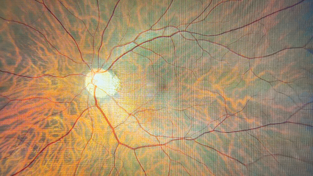
The (Unknown) Power of Retinal Imaging
Researchers use retinal images to predict risk of eye and systemic diseases
Many previous studies have searched for genes associated with overall retinal health, but neglected to investigate the role of the different cell layers that make up the retina. Now, researchers at Harvard Medical School and the Broad Institute of MIT and Harvard, have paired retinal imaging, genetics, and big data to estimate how likely a person is to develop eye – and systemic – diseases in the future (1).
“Our research sought to differentiate the nine retinal layers, all of which reflect distinct cell types and play a contrasting functional role, including different types of neuronal cells, vascular cells, and epithelial cells,” says Maryam Zekavat, co-author and researcher at the Department of Ophthalmology, Massachusetts Eye and Ear, Harvard Medical School, Boston.
The researchers analyzed data from around 50,000 participants, who all first received optical coherence tomography (OCT) imaging of the retina, genotyping, and baseline measurements of health in 2010 and were then followed for disease development over an average of 10 years. The phenome- and genome-wide analyses led to the discovery of associations between the thinning of different retinal layers and increased risk across all disease categories. “We identified multiple inherited genetic loci and acquired systemic cardio-metabolic-pulmonary conditions associated with thinner retinal layers, and identified retinal layers wherein thinning is predictive of future ocular and systemic conditions,” Zekavat adds.
These analyses also helped assess the biological pathways related to the different retinal layers. Could this be the start of targeted therapies for ocular conditions linked to retinal nerve fiber layer and photoreceptor segment thinning? Though that answer remains to be seen, the findings look to be a win for a new wave of preventative medicine. “There is a lot more information we can gain from our retina images than we originally thought was possible,” says Nazlee Zebardast, HMS Assistant Professor of Ophthalmology and Director of Glaucoma Imaging at Mass Eye and Ear. “This could potentially help with disease prevention. If we know from someone’s retinal image that they are at high risk of developing glaucoma or cardiovascular disease in the future, we could refer them for follow-up screening or preventative treatment.”
The team has since developed the Ocular Knowledge Portal to showcase their findings.
This article first appeared in The Ophthalmologist.
Reference
- SM Zekavat et al., “Phenome- and genome-wide analyses of retinal optical coherence tomography images identify links between ocular and systemic health,” Science Translational Medicine., 16, 4517 (2024). PMID: 38266105.
The New Optometrist Newsletter
Permission Statement
By opting-in, you agree to receive email communications from The New Optometrist. You will stay up-to-date with optometry content, news, events and sponsors information.
You can view our privacy policy here
Most Popular
Sign up to The New Optometrist Updates
Permission Statement
By opting-in, you agree to receive email communications from The New Optometrist. You will stay up-to-date with optometry content, news, events and sponsors information.
You can view our privacy policy here
Sign up to The New Optometrist Updates
Permission Statement
By opting-in, you agree to receive email communications from The New Optometrist. You will stay up-to-date with optometry content, news, events and sponsors information.
You can view our privacy policy here


